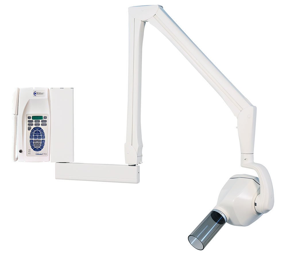
X-ray unit
Leadex70
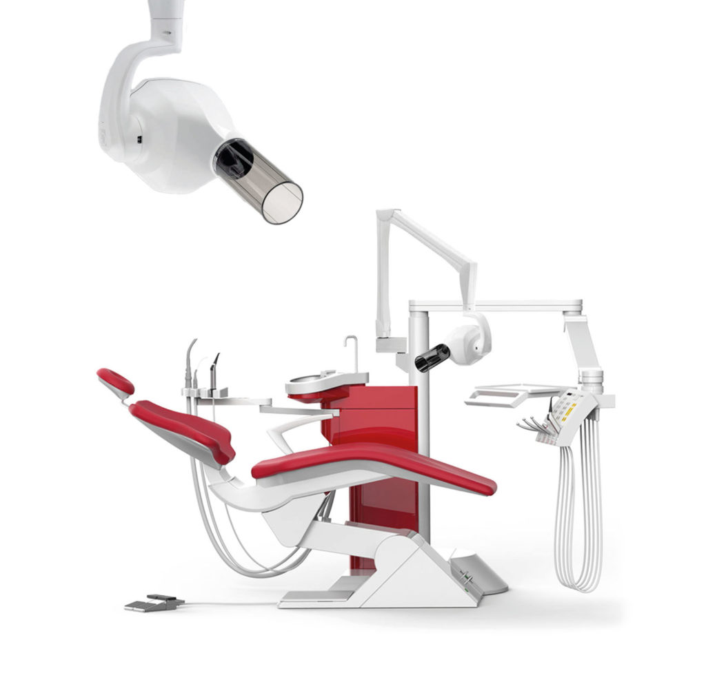
Leadex70
The Leadex70 is designed for all intraoral dental radiology requirements in the modern dental practice. Integrated directly on your Ritter dental unit, the Leadex70 is a flexible, fast and safe solution to produce an excellent small X-ray image. Compatible with standard films and digital X-ray sensors, it guarantees sharp and high-contrast images at an attractive price. The control unit is easy and intuitive to use. It can be mounted directly on the wall in the treatment room or in an adjacent room (cable from the unit in the floor). The X-ray process is performed via the control panel or via the supplied remote control unit.
TECHNICAL DATA
X-RAY DEVELOPER
GIRARDELLI X-3D
The unbeatable classic in the
X-ray film development
The GIRARDELLI X-3D immersion x-ray processor with integrated daylight attachment fulfils all present requirements of a modern and efficient developing automat:
Integrated daylight attachment
Processing under daylight conditions. A darkroom is not necessary.
Auto-activator
When pressing the start button, the transport shaft and the dryer-blower are now operative. A the end of the development process, the appliance will be switched off by pressing the start button again.
Electronical bath heating
The electronically regulated bath heating guarantees a steady temperature at all times. This way an optimal picture quality and life span of the GIRARDELLI developing concentrate (up to 12 weeks per batch) is achieved.
Automatic film drying
Very powerful dryer-blower for fast and efficient film drying.

TABLE DARK ROOM
GIRARDELLI X-1
The GIRARDELLI X-1 table darkroom meets all today’s requirements of an efficient hand-developed device.
Technical data
Dimensions:
46 x 32 x 30 cm (W x D x H)
Contents of the cups:
4 x 0.225 litres
Weight:
approx. 2.3 kg
Standard equipment:
detachable red light lens, 3 film holders for single films, 4 x 0.225 litre plastic cups with screw lid closure
- Each part used is extremely stable and chemically resistant
- Minimal service costs over many years
- Ideal cup size
- Allows the development of intraoral film sizes
- Even occlusal films can be developed
- Minimal maintenance
- Only short cleaning of the cups necessary when changing the used GIRARDELLI powder development concentrate.
- Simplest operation
- The film holders can be loaded and the films can be developed via the manual interventions.
- Slanted top allows for excellent visibility

DISINFECTION MACHINE
FOR THE HANDS
GIRARDELLI DA-1
The purpose of the GIRARDELLI DA-1 disinfection automat is the hygienic and surgical hand disinfection in the entire medical- and food-industry.
Efficient and sparing.

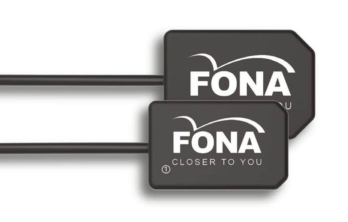
StarX PRO
Intraoral sensor
StarX PRO sensors are the new FONA Intraoral sensors builtwith the latest technologies to deliver perfect images at every shot. Thanks to the USB direct connection, StarX PRO provides brilliant and real-time images with a simple and effective plug and play solution.
Engineered to be a flexible and a user-friendly solution, StarX PRO come in size 1 and 2. With round edges and ergonomic design, it enhances the patient comfort and ease the positioning procedures made by the operator.
TECHNICAL DATA
• Sensor sizes: 1, 2
• Measured resolution: 20 lp/mm
• Theoretical resolution: 25 lp/mm
• Technology: CMOS with CSI scintillator
• Pixel Size: 20 _m
• Sensor Shape: Round edges for patient comfort
• Sensor activation: Plug & play solution, always ready to acquire
• Sensor cable length: 2 m
• Connectivity: USB 2.0
• Dimensions: Size 1: 35,4×28,1×5,3mm; size 2: 39x25x5,6mm
• Active area dimensions: Size 1: 20x30mm, size 2: 26x34mm
• Software: OrisWin DG Suite
• Operating System Support: Windows 10, Windows 8, Windows 7
• Licenses: Up to 10 workstations running simultaneously with one license
• DICOM support: Yes
• Compatibility with Intraoral Generators: Yes, both AC and DC technology, 60-70kV
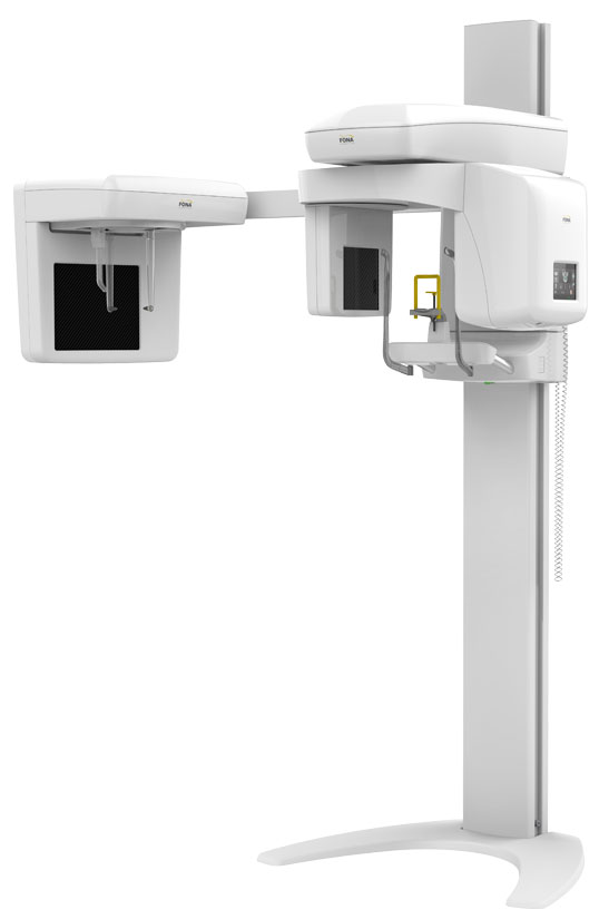
Stellaris
Panoramic dental X-ray
Stellaris 3D is a complete and compact fully upgradeable 3D CBCT, Panoramic and Cephalometric system, which covers an entire range of applications – from endodontics, periodontology, surgery, implantology to orthodontics – everything you need in your daily practice.
TECHNICAL DATA
• 3D Programs: 23 programs: adult full, child full, adult and child upper and lower jaw, …
• Volume size 3D: 8 volumes: 5×6.5, 7×6.5, 6×10, 7×10, 8×6.5, 8×10, 10×6.5, 10×10 (in cm)
• Voxel size: 80 μm, 190 μm
• Exposure time 3D: 12.4 – 13 s (STD mode) | 16.1 – 16.9 s (HD mode)
• Panoramic Programs: 9 programs: adult pano, child pano, dentition, Panoramic bitewing, …
• Pixel Size: 99 μm
• Exposure time 2D: 4.6 – 14.2 s (STD mode) | 3.2 – 9.9 s (ECO mode)
• Ceph Programs: LL 30×24, LL 18×24, AP, PA, Carpus
• Ceph Image Size: 240×300 mm, 240×180 mm
• Exposure time Ceph: 0.2 – 4.0 s
• Patient Sizes: small, medium, large and extra-large
• Patient position: Standing or sitting
• Wheelchair accessible: Yes
• Positioning: against mirror, 3 aiming light beams, temporal support
• Weight: 109 kg without ceph, 137 kg with ceph
• Footprint: without arm 97.5 cm x 117.5 cm x 222 cm; with arm 189 cm x 117.5 cm x 222 cm
• Fixation: wall fixation and floor fixation with baseplate
• Power supply: 230 V ± 10%, 115 V ± 10%, 50/60 Hz
• Computing system: Included, with preinstalled reconstruction and viewing software and patient database. Monitor not included
• Connectivity: Stellaris 3D/3D Ceph: 2 ethernet cables
Stellaris 2D/2D Ceph: 2 ethernet cables + USB 3.0 adapter

ONE
Intraoral sensor
ONE is an intraoral sensor featuring CMOS technology. Its ergonomics and design have been designed to adapt perfectly to your practice’s needs. The smooth corners and rounded sides of this device allow your patient to be positioned with the utmost ease. The sleek sensor and discrete cable make for optimum comfort in the mouth.

TECHNICAL DATA
• Sensor: CMOS
• Dimensions: 39 x 25 x 5 mm
• Sensitive surface: 30 x 20 mm (600 mm2)
• Surface in pixels: 1500 x 1000 pixels
• Sensor cable length: 3 m
• Grey levels: 14 bits (16384 grey levels)
• Connection Standard USB: USB 2.0 High-Speed (480Mbit/s) and USB 3.0
CR2 Table-Top Unit
Intraoral Imaging Plate Reader
4 plate sizes for a scanner suitable for all dental practices. The Owandy-CR2 can be used in endodontics, in prosthetic and implant surgery, in periodontics, but also for caries diagnostics. Different plate sizes are available to best meet your requirements (bitewing, retroalveolar, posterior, etc.) and your patients.

TECHNICAL DATA
• Technology: Re-usable imaging plate (PSP: Photostimulable Phosphor plate)
• Resolution: 34 pixel/mm – 16 Bits
• Pixel size: 30 μm
• Scan time: 4 – 9 s
• Connection: USB 2.0 & USB 3.0
• Ethernet: 10/100/1000 Mbps
• Dimensions: 286 x 151 x 104 mm
• Weight: 4,25 kg
• External power supply: 100-240 V, 50/60Hz, 60W
• Plate size selection: Automatic detection
• System standby mode: Automatic function
• Operating system: Windows 7, 8, 10 (32-64 Bits)
Plate size selection:
• Size 0: 31 x 22mm, 1024 x 726 pixels
• Size 1: 40 x 24mm, 1321 x 792 pixels
• Size 2: 41 x 31mm, 1354 x 1024 pixels
• Size 3: 54 x 27mm, 1783 x 891 pixels
RX Portable
Portable X-ray system
Weighing only 1.8kg, the Owandy-RX Portable is one of the lightest dental X-ray devices on the market. It is therefore easy to handle and position. In your practice, whether in the chair or in the surgery room, our portable X-ray generator gives you total autonomy in your movements.
Take up to 300 images on a single charge!

TECHNICAL DATA
• X-ray tube: CEI
• kV/mA: 70kV / 3mA
• Focal spot: 0.3 mm
• Total filtration: 1.5mm Al
• Battery charger input voltage: 100-240V / 50~60Hz
• EBattery voltage: 24.2V
• Weight: 1.8 kg
• Options Backscatter protection; Rectangular collimator (30x40mm, 20×30 mm)

RX AC/DC
X-ray generator
Owandy-RX-AC-DC provide very precise exposure times with guaranteed control thanks to microprocessor controlled timer. Keep your patients safe with HF technology that reduces x-ray doses by eliminating harmful soft rays. Activates automatic exposure time compensation, regardless of any fluctuations in the mains supply. The flexible arm allows the Owandy-RX to be handled effortlessly, with accurate positioning.

TECHNICAL DATA
• Category: Class I, type B (CEI EN60601-1:2007)
• Supply voltage: 115/220/230 V – single phase 50/60 Hz (AC); 115/230 V – single phase 50/60 Hz (DC)
• Power input: 0,8 kVA (AC); 1,4 kVA (DC)
• High voltage: 70 kV (AC); 60 à 70 kV (DC)
• Anode current: 8 mA (AC); 4 à 8 mA (DC)
• Total filtration: Equivalent to 2 mm Al. à 70 kV
• Leakage radiation: < 0,25 mGy/h
• Technology: Alternating current (AC); Direct current (DC)
• Timer: from 0.08 to 3.2 secs AC); from 0.02 to 3.2 secs (DC)
• Head weight: 9 kg (AC); 5,5 kg (DC)
• Total weight: 23 kg (AC); 19,5 kg (DC)
• Arm extensions: 41 cm / 82,5 cm / 110 cm
• Options: Mobile version (AC); Mobile version, Wireless (DC)
• Accessories: Long cone (31cm) / Long rectangular cone (31cm)

I-Max
Dental Panoramic Imaging System
Multifunctional dental panoramic for enhanced diagnostics. The I-Max is not only the lightest dental panoramic scanner on the market, it’s also the model with the best price/performance ratio. I-Max is a 4-in-1 Dental Cone Beam designed to enable more accurate diagnosis, treatment planning, and better outcomes for patients. Our dental panoramic seamlessly integrates into the digital workflow. From imaging to guided surgery, it ensures the highest level of precision and safety. I-Max panoramic adapts to you and your current needs. It allows you to switch from traditional 2D to a 2D/3D Cone Beam version whenever you want.

TECHNICAL DATA
• Category: II B / CE0051
• Power supply: 110-120 V, 220-240 V at 50/60Hz
• Anode voltage: 70 kVp (I-Max); ±8% 86 kVp ±8% (I-Max Ceph, I-Max 3D, I-Max Ceph 3D)
• Anode current: 7,1 mA ±10% (I-Max); 12,5 mA ±10% (I-Max Ceph, I-Max 3D, I-Max Ceph 3D)
• SID (Source to Image Distance): 50 cm 52 cm (2D), 165 cm (Ceph)
• Total weight: 62 Kg (I-Max); 120 Kg (I-Max Ceph); 66 Kg (I-Max 3D); 125 Kg (I-Max Ceph 3D)
• Inherent filtration: 2 mm Al eq. @ 70kVp (I-Max); ≥ 2,5 mm Al. eq @ 86kVp (I-Max Ceph, I-Max 3D, I-Max Ceph 3D)
• HF generator: Constant potential (DC)
• X-Ray focal spot: 0,5 mm EN 60336
• Connection: LAN, Ethernet (without dedicated PC) (I-Max); LAN Ethernet (dedicated PC) (I-Max Ceph, I-Max 3D, I-Max Ceph 3D)
• Voxel: n/a (I-Max); n/a (I-Max Ceph); 87,5 μm (min. cross-section depth) (I-Max 3D, I-Max Ceph 3D)
• Exposure time: 2,44 to 14,4 s (PAN adult/child) (I-Max, I-Max Ceph); 3,20 to 14,4 s (2D), 10,8 to 11,2 s (3D)
• Panoramic: I-Max, I-Max Ceph, I-Max 3D, I-Max Ceph 3D
• Cephalometric: I-Max Ceph, I-Max Ceph 3D
• 3D cone beam: I-Max 3D, I-Max Ceph 3D
• FOV: 12×10, 9×9, 9×5, 5×5 (I-Max 3D, I-Max Ceph 3D)
• Fitting type: Wall (I-Max, I-Max 3D); Floor + Wall (I-Max Ceph, I-Max Ceph 3D)
• Options: Floor base plate, Wall column (I-Max, I-Max 3D); Floor base plate (I-Max Ceph, I-Max Ceph 3D)
Versions:
• I-Max digital pan 2D
• I-Max digital pan 2D, with ceph arm
• I-Max 3D, wall mount compact unit
• I-Max 3D with Ceph

Ceph Analysis
Orthodontic Software
Stop losing time… Use Artificial Intelligence to automate your work with Ceph Analysis.
The automatic tracing feature is powered by a high-tech industry proven Artificial Intelligence engine. A.I. automatically finds soft and hard tissue landmarks, planes and silhouettes on lateral or PA radiographs within seconds. Analyses and patient’s images can be superimposed automatically or manually. You can manage how analyses are displayed, automate report generation, visualize growth and treatment effects.

FEATURES
Image Analysis & Overlay:
Patient analyses and images can be automatically or manually overlaid. You can manage the display of analyses, automate report generation, and visualize growth and treatment effects.
Visualize Expectations with Growth Projection:
The skull growth projection will increase the skull trace from the current skeletal age to the chosen skeletal age. Projections up to maturity are possible.
Treatment Plans Based on Predictions:
VTO and STO will assist in creating treatment plans. By simulating orthodontic and/or surgical treatment, a detailed treatment plan can be created. Considering different tissue stiffness levels, excellent prediction results are achieved.
Over 200 Types of Analyses:
Choose from over 200 existing analysis types or create your own. Become your own architect!
Manage Patient Data with Powerful and Secure Tools:
Looking for your patient’s data? Documents are carefully organized by treatment stage. Data can be encrypted to enhance security.
Cam HD
Inraoral camera
Its “focus-free” concept delivers enhanced field depth and high quality images. Requiring no adjustments at all, this unit delivers perfectly clear images, from a full smile to just one tooth – and will save you time, too! The new HD Owandy-Cam will assist you in your everyday work, enabling you to better communicate with patients. An image is worth a thousand words! Explain clinical procedures to your patients more easily! Your patients will feel more involved and have greater trust in the process.

TECHNICAL DATA
• Sensor: High sensitivity 1⁄4 ” Sony CCD
• Resolution: 50 images / sec
• Lighting: 8 LEDs
• Adjustment: Focus free, large field depth
• View angle: 105°
• USB cable length: 2.5m
• Handpiece size: (L x W x H) 205 x 28 x 24mm
• Thickness of intraoral part: 11mm
• Handset weight: 55g
• Image setter: Touch button

Port-X III
Portable X-ray system
coming soon

TECHNICAL DATA
coming soon
